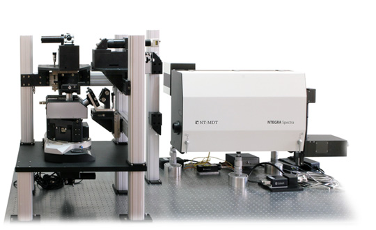
 |
NTEGRA Spectra II
Versatile automated AFM-Raman, SNOM and TERS system.
Part number:
Supplier:
NT-MDT Spectrum InstrumentsDescription
- High-performance versatile AFM
- Optical access from top, side and bottom optimized for Raman, TERS and SNOM
- Flexible optical design providing any combination of excitation/collection configurations
- Automated AFM laser, probe and photodiode alignment
- User-friendly change of wavelength of AFM registration system laser and photodiode
- Easy and user-friendly change of objectives
- Integration with IR s-SNOM (optional)
Since 1998 NT-MDT has been successfully integrating AFM with optical microscopy and spectroscopy techniques. More than 30 basic and advanced AFM modes including HybriD ModeTM are supported providing extensive information about the sample surface physical properties. Integration of AFM with confocal Raman/fluorescence microscopy provide the widest range of additional information about the sample.
Simultaneously measured AFM and Raman maps of exactly the same sample area provide complementary information about sample physical properties (AFM) and chemical composition (Raman).
NTEGRA Spectra II with the help of Tip Enhanced Raman Scattering (TERS) allows carrying out spectroscopy/microscopy with nanometer scale resolution. Specially prepared AFM probes (nanoantennas) can be used for TERS to enhance and localize light at the nanometer scale area near the tip apex.
Such nanoantennas act as a “nano-source” of light giving possibility of optical imaging with resolution less than a diffraction limit (up to ~ 10 nm). Scanning near-field optical microscopy (SNOM) is another approach to obtain optical and spectroscopy images of optically active samples with resolution limited by probe aperture size (~ 100 nm).