 |
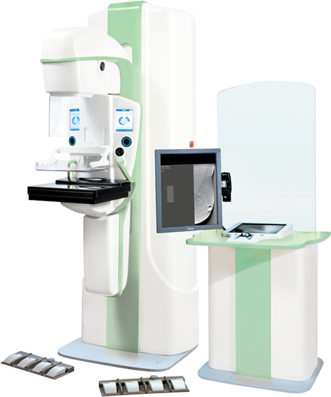
Mammo-5MT Digital Breast Tomosynthesis System
The Mammo-5MT X-ray DBT unit provides high image quality that is vital for precise diagnostics of breast diseases, including early detection of breast cancer even in cases of high-density breast tissue.
Part number:
Supplier:
Medical Technologies Ltd.Description
The unit is intended for acquisition of a series of high-resolution breast images for 3D reconstruction. Its advantages include faster image acquisition, lower patient exposure dose and lower breast compression force.
Mammo-5MT Digital Breast Tomosynthesis System
The DBT unit captures a series of high quality tomographic images and, using an optimized 3D reconstruction algorithm, creates a 3D image of the breast, thus minimizing the need for additional tests like spot mammography or other imaging techniques. Another benefit of Breast Tomo imaging is a lower overall patient exposure to X-ray dose levels.
During the exposure the X-ray tube moves in an arc around the breast to produce up to 25 images taken at different angles. A special algorithm built into the acquisition workstation software processes the digital images captured and renders a 3D image of the breast. To achieve the optimum level of detail, a radiologist may adjust the slice thickness during the 3D imaging process.
A high resolution digital X-ray detector, based on CMOS technology, is specifically designed to capture tiny artifacts as may be evident in breast imaging. The detector ensures high speed of image acquisition and operates at a wider temperature range compared to detectors based on a-Se (amorphous selenium).
Digital X-ray detector
Digital X-ray detector
The CMOS-based X-ray detector allows for acquisition of high-resolution, low-noise images with higher dynamic range, which results in better recognition of both micro-calcification and low-contrast objects on images.
Specially configured review workstation software allows radiologists to view volumetric images as well as any individual reconstructed slice, removing layers of overlying and underlying tissues to more accurately identify the spatial position of an area of interest in a breast.